At a glance:
- High environmental temperatures decrease endurance performance.
- Oxygen delivery to the working muscles is reduced via redirection of total available blood.
- Blood plasma volume is reduced via sweating, resulting in a lower stroke volume (the amount of blood ejected from the heart with each beat).
- At a given intensity, more carbohydrates are used for fuel in hotter conditions, resulting in faster glycogen depletion.
For a simplified version, check out the corresponding YouTube video for this article.
It's well documented that high environmental temperatures reduce endurance performance. When comparing conditions at or above 30°C to cool conditions, the average reduction in 40km time trial power output across multiple studies is 15% [1]. This is a massive difference. To help visualize this difference, it's equivalent to an individual who can average 350W for one hour, only holding 298W. This article will focus on explaining the three biggest physiological reasons why endurance performance suffers in the heat. This is the first step towards understanding if we can adapt to heat, and more importantly, designing a heat acclimatisation protocol.
Importance of Maintaining Body Temperature
Your body needs to maintain a stable internal environment to function. For example, your body will tightly regulate blood pressure, blood glucose, water levels, and pH. This is known as homeostasis.
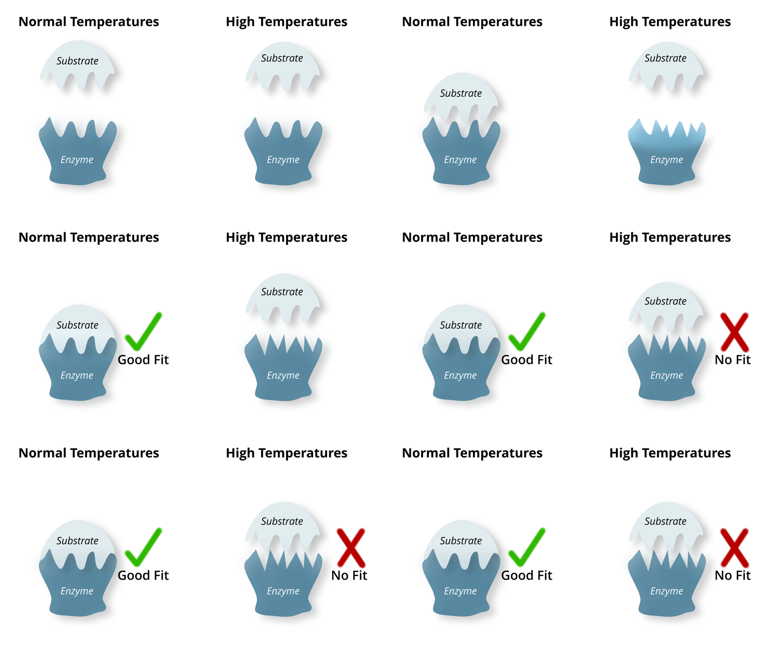
An important component of homeostasis is the regulation of core body temperature. In humans, the core body temperature must be kept in a narrow range around 37°C [2]. This is crucial for allowing enzymes and cells to carry out vital biological processes. Even a slight change in core body temperature can cause enzymes to denature (change shape), meaning they are no longer functional. This is because substrates can no longer fit into their active site (see Figure 1). If core body temperature continues to rise, this can lead to stroke, seizures, and organ failure [3]. Therefore, it is not surprising why your body will always prioritise maintaining core body temperature over power output whilst cycling.
Increased Cardiovascular Load
When you exercise in the heat, the very first system that gets overloaded isn't your muscles, it's your cardiovascular system. One of the ways your body cools you down is to redirect blood away from your working muscles.
The process starts when central thermoreceptors detect a change in core body temperature. Unlike peripheral thermoreceptors which are concerned with surface skin/environmental temperature, central thermoreceptors are concerned with core body temperature. Their location reflects this function, being present in the hypothalamus (brain), spinal cord, and internal organs [4]. The central thermoreceptors send their temperature signals to the hypothalamus. The hypothalamus is your body's thermoregulatory centre, acting like a thermostat to monitor and maintain core body temperature. Once the signals are received by the hypothalamus, it activates the negative feedback mechanisms to cool the body down.
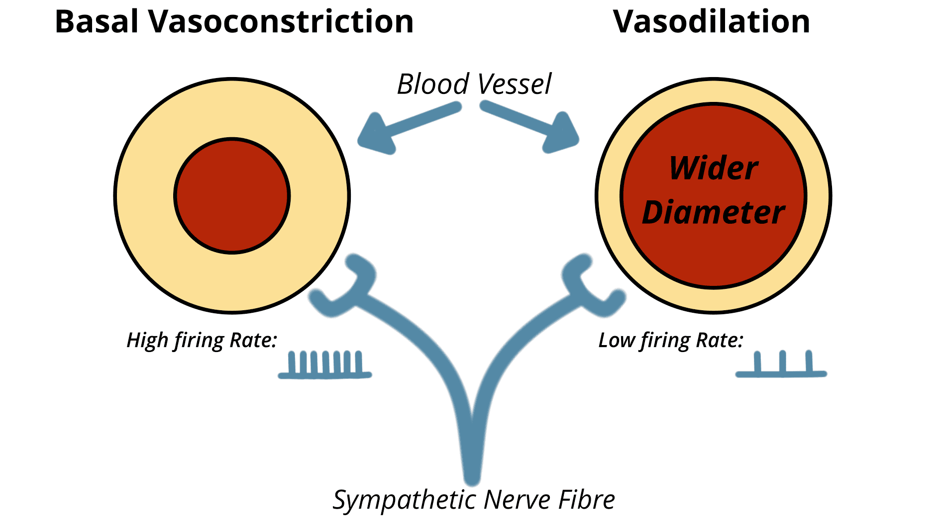
One of the mechanisms the body uses to reduce temperature works by increasing the amount of blood near the skin's surface. Under normal conditions, the skin surface blood vessels maintain their diameter by a partial level of vasoconstriction (narrowing of the blood vessel). This level is maintained by a constant firing rate from sympathetic nerves. When temperature rises, the hypothalamus decreases the sympathetic tone and therefore reduces the firing rate of these nerves (see Figure 2). This results in vasodilation (widening of the blood vessels), allowing more blood to flow through them. In addition to this, the hypothalamus actively dilates blood vessels by sending out the neurotransmitter and vasodilator: ACh (acetylcholine). Now with much wider blood vessels near the surface layer of skin, there is more blood that can contribute to heat loss via radiation and convection. More blood is being directed away from the working muscles. Put simply, there is now less oxygen being delivered to the working muscles, directly reducing performance. Your heart is essentially being asked to perform two jobs: maintain power output and reduce temperature. The strain on the cardiovascular system has increased drastically. This strain is amplified further in our next reason, where blood is once again moved away from the working muscle tissue.
Dehydration or Reduced Plasma Volume
A key component of endurance performance is blood plasma volume. The increased sweat rate in the heat requires more water to be pulled out of the blood, thus lowering blood plasma levels, impairing performance.
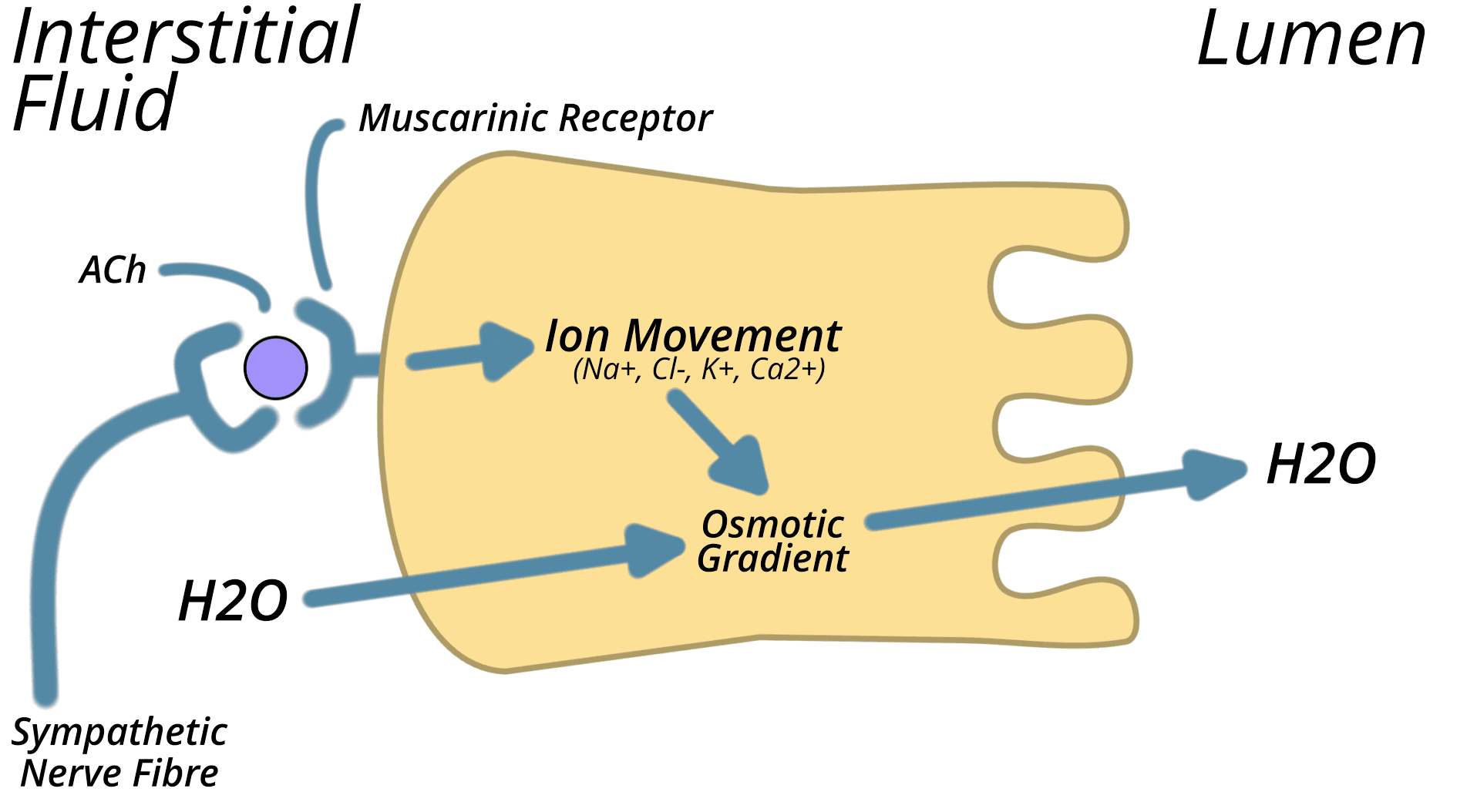
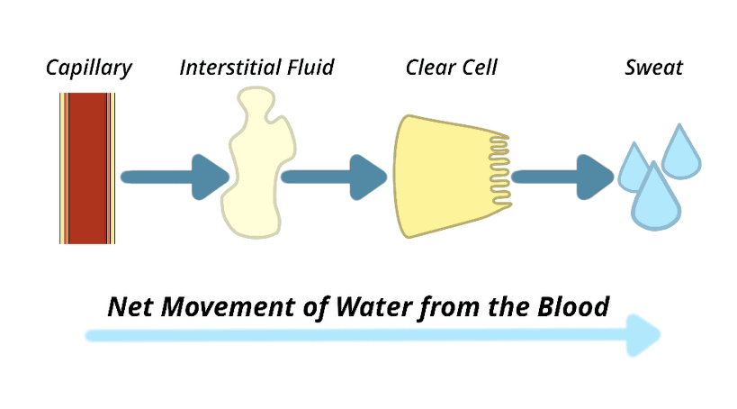
During heat stress, more blood is directed to the sweat glands. Blood plasma is filtered out of the capillaries (small blood vessels), forming interstitial fluid around the sweat gland cells. Sweat glands are made up of three types of cells: clear, dark, and myoepithelial [5]. We are most interested in clear cells, as they are largely responsible for sweat production and secretion. As mentioned in the previous section, under heat stress, the hypothalamus increases the release of ACh from sympathetic nerve fibres. ACh binds to muscarinic receptors on the basolateral membrane of clear cells which initiates ion movement (see Figure 3). This ion movement results in the creation of an osmotic gradient, that forces water to move from the interstitial fluid, through the clear cell, into the sweat gland lumen where it can be secreted to the skin surface (see Figure 4).
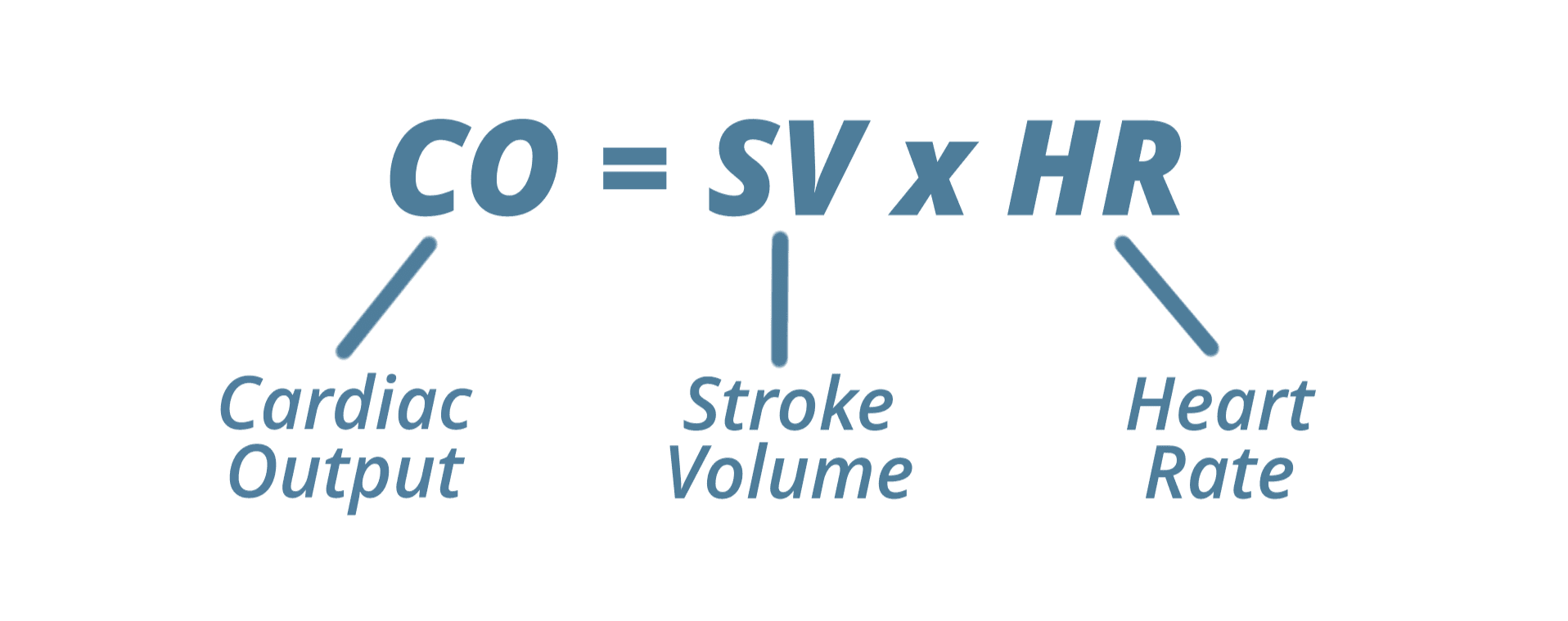
This reduction in plasma from the blood results in a loss of stroke volume, which is the amount of blood ejected from the heart with each beat. This can be explained by the Frank-Starling law, which is the observation that stroke volume increases as preload increases (amount of stretch in the heart muscle fibres before they contract) [6]. Preload increases with venous return (the amount of blood returning to the heart). With reduced plasma volume, the venous return is lowered, therefore reducing preload, and consequently lowering stroke volume. However, your muscles still require a certain volume of blood per minute. With a lower stroke volume, the only way to maintain the same cardiac output (the amount of blood ejected from the heart per minute) is to increase heart rate (see Figure 5).
A higher heart rate at the same workload (power output) will result in an earlier onset of fatigue. This is due to two reasons. Firstly, a higher heart rate will require more oxygen for the myocardium (heart muscle). Secondly, a higher heart rate limits your cardiac reserve (the number of beats you have before you reach max heart rate). When heart rate is elevated at a given intensity, there is less capacity to further increase cardiac output as exercise intensity rises. Along with water loss, sweat also loses sodium and potassium which are essential for proper nerve and muscle function. Additionally, sweat glands run almost entirely on glucose, which adds to our next driver of reduced performance [5].
Increased Carbohydrate Utilisation
Heat stress shifts metabolism towards a greater reliance on carbohydrates, and away from fats as a fuel source [7]. There are a few reasons for this, but the most potent is likely the increase of epinephrine (also known as adrenaline) that exercising at higher temperatures induces.
As mentioned previously, your body is preventing some serious side effects whilst exercising in the heat (stroke, organ failure). This perceived potential danger activates your sympathetic nervous system (fight or flight response). Once activated, the hypothalamus sends signals to the adrenal glands (located above each kidney) to produce and release the hormone epinephrine. Epinephrine travels in the blood, binding to receptors on liver and muscle cells (see Figure 6). This results in a phosphorylation cascade that results in the addition of a phosphate group (P) to the enzymes: glycogen phosphorylase & glycogen synthase. The addition of a P to glycogen phosphorylase causes it to become more active. Glycogen phosphorylase breaks down glycogen into glucose. Therefore, more glucose is readily available in the muscle cell. The opposite is true for glycogen synthase; the addition of a P deactivates it. When active, glycogen synthase converts glucose into the storage molecule, glycogen. Therefore, even more glucose is readily available in the muscle cell to be used for energy.
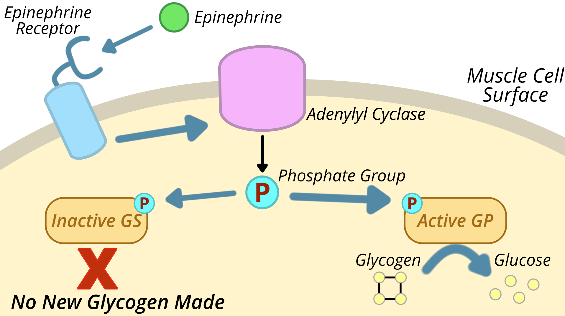
Together, these mechanisms shift the body toward greater carbohydrate usage, resulting in greater glycogen depletion at the same power output and therefore earlier time to exhaustion.
Summary
These are the three main central limitations to performance loss in the heat: increased cardiovascular strain, dehydration, and faster glycogen depletion. Future posts will investigate how much performance increase we can expect by undertaking heat training, what the adaptations are that occur during heat training, and how to devise the ideal heat training protocol for your own personal goals.
References
- Junge, N., Jørgensen, R., Flouris, A. D., & Nybo, L. (2016). Prolonged self-paced exercise in the heat: environmental factors affecting performance. Temperature, 3(4), 539-548. https://doi.org/10.1080/23328940.2016.1216257
- Yousef H, Ramezanpour Ahangar E, Varacallo MA. Physiology, Thermal Regulation. In: StatPearls [Internet]. Treasure Island (FL): StatPearls Publishing; 2025. https://www.ncbi.nlm.nih.gov/books/NBK499843/
- Marchand, M., & Gin, K. (2021). The Cardiovascular System in Heat Stroke. CJC Open, 4(2), 158-163. https://doi.org/10.1016/j.cjco.2021.10.002
- Osilla EV, Marsidi JL, Shumway KR, et al. Physiology, Temperature Regulation. In: StatPearls [Internet]. Treasure Island (FL): StatPearls Publishing; 2025. https://www.ncbi.nlm.nih.gov/books/NBK507838/
- Baker L. B. (2019). Physiology of sweat gland function: The roles of sweating and sweat composition in human health. Temperature, 6(3), 211-259. https://doi.org/10.1080/23328940.2019.1632145
- Delicce, A.V., & Makaryus, A.N. (2023). Physiology, Frank Starling Law. In: StatPearls [Internet]. StatPearls Publishing. https://www.ncbi.nlm.nih.gov/books/NBK470295/
- Febbraio, M. A., Snow, R. J., Hargreaves, M., Stathis, C. G., Martin, I. K., & Carey, M. F. (1994). Muscle metabolism during exercise and heat stress in trained men: effect of acclimation. Journal of Applied Physiology, 76(2), 589-597. https://doi.org/10.1152/jappl.1994.76.2.589